Research/ Medical Image Processing
Introduction
Advances in science and technology have made image-based diagnosis using X-ray, CT, and MRI commonplace in clinical practice. Computer analysis of medical images supports physicians in diagnosis. Here we describe our research on brain MRI analysis and ultrasound image analysis.
Brain MRI
Brain MRI is used to assess brain condition. We focus on T1-weighted images employed for morphological diagnosis. As shown in Figure 1, three brain tissue types can be distinguished in T1-weighted images.
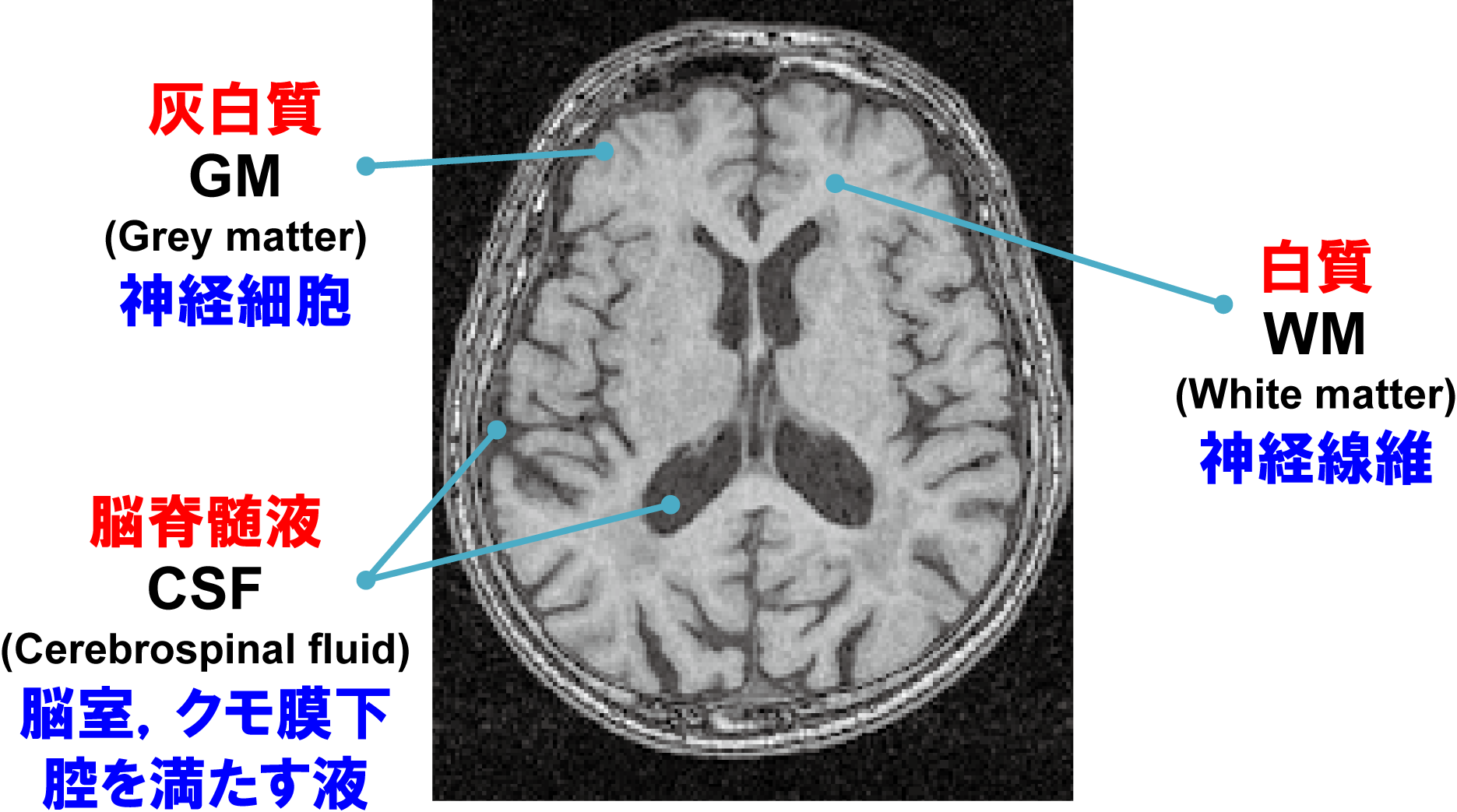
Figure 1: T1-weighted brain image and brain tissues
Age-Related Atrophy of Brain Morphology
It is known that brain morphology atrophies with aging. Figure 2 shows graphs of volume changes in each brain tissue with age, together with actual T1-weighted images.
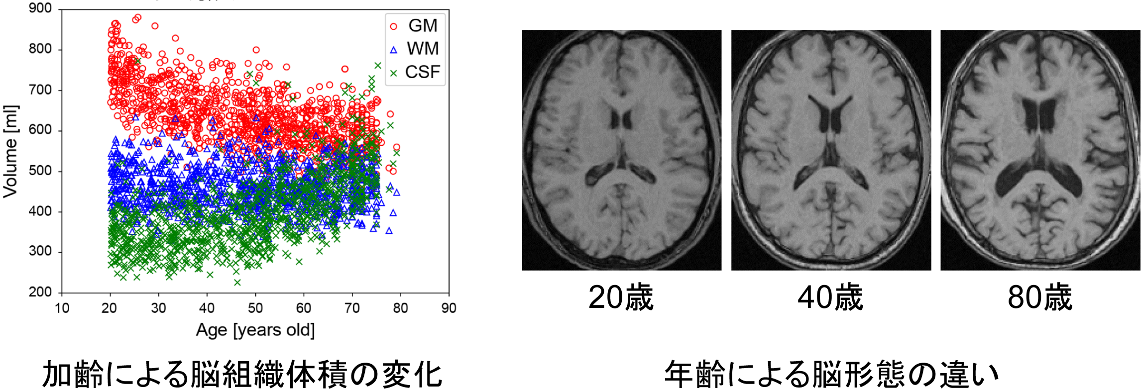
Figure 2: Age-related volume changes in brain tissues and morphological differences across ages
Age Estimation
Our laboratory exploits the strong relationship between morphological change and age to estimate chronological age from T1-weighted images. With state-of-the-art 3D CNN-based methods, estimation error has been reduced to approximately three years.
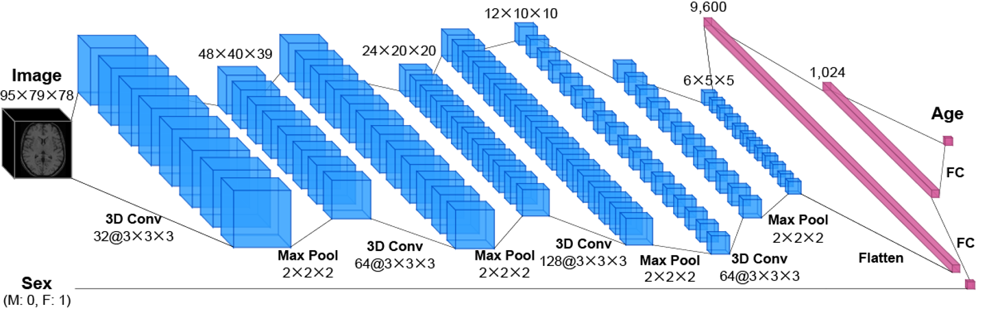
Figure 3: Age estimation network using a 3D CNN
Diagnostic Support for Alzheimer's Disease
We pursue early detection of Alzheimer's disease (AD), one of the major brain disorders. For details, see reference [3].
Ultrasound Images
Ultrasound diagnosis offers advantages beyond compact equipment: it is non-invasive and places minimal burden on patients. Our laboratory investigates methods to reconstruct three-dimensional volume data from ultrasound images.
Ultrasound Probe–Camera System
As shown in Figure 4, we developed an ultrasound probe–camera system by mounting a camera on an ultrasound probe. For methodological details, see reference [4].
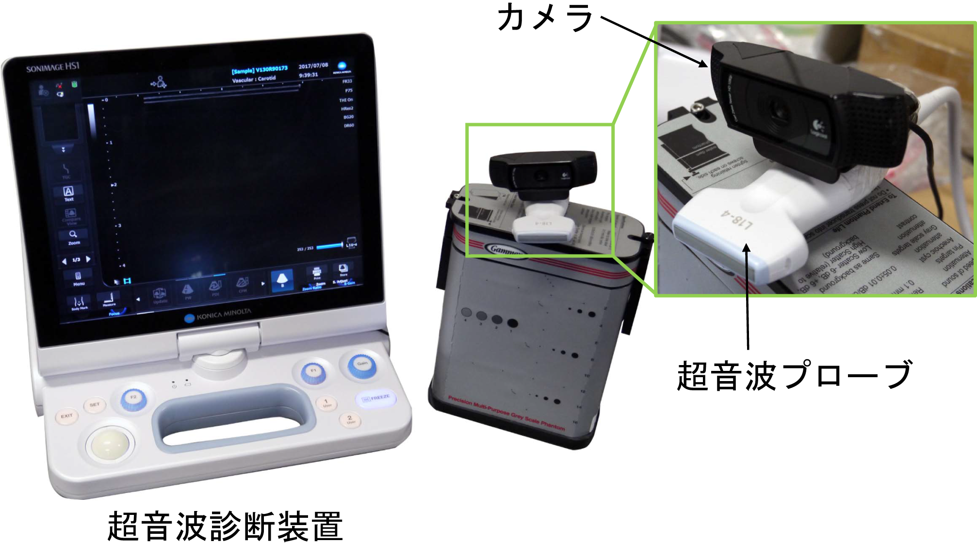
Figure 4: Ultrasound probe–camera system
3D Ultrasound Image Reconstruction
Research is underway to reconstruct 3D ultrasound volumes from ultrasound images alone using deep learning. Our work to date is described in reference [5].
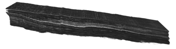
Figure 5: 3D ultrasound image of the lower leg
3D Retinal Image Analysis
Many ocular diseases such as glaucoma alter retinal structure. We investigate deep-learning methods to measure retinal layer thickness. Our work to date is described in references [6] and [7].
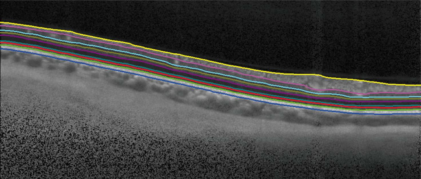
Figure 6: 3D retinal image analysis (retinal layer boundary detection)
Summary
We have briefly introduced medical image processing research in our laboratory. Medical image processing is an interdisciplinary field requiring knowledge of both medicine and engineering.
References
- C. Kondo et al., "Age estimation method using brain local features for T1-weighted images," Proc. Annual Int'l Conf. IEEE Engineering in Medicine and Biology Society, pp. 666-669, August 2015.
- M. Ueda et al., "An age estimation method using 3D-CNN from brain MRI images," Proc. Int'l Symp. Biomedical Imaging, pp. 380-383, April 2019.
- 遠藤ほか, "畳み込みニューラルネットワークと認知機能テストを用いたアルツハイマー病鑑別," 第24回 画像の認識・理解シンポジウム, July 2021.
- K. Ito et al., "A probe-camera system for 3D ultrasound image reconstruction," Proc. Int'l Workshop on Point-of-Care Ultrasound, pp. 129-137, September 2017.
- K. Miura et al., "Localizing 2D ultrasound probe from ultrasound image sequences using deep learning for volume reconstruction," Proc. Advances in Simplifying Medical UltraSound (MICCAI 2020 Workshops), pp. 97-105, October 2020.
- T. Konno et al., "Retinal layer segmentation from OCT images using 2D-3D hybrid network with multi-scale loss and refinement module," Proc. Int'l Symp. Biomedical Imaging, April 2023.
- T. Konno et al., "Retinal layer segmentation using 1D+2D U-Net from OCT images," Proc. Int'l Symp. Biomedical Imaging, May 2024.